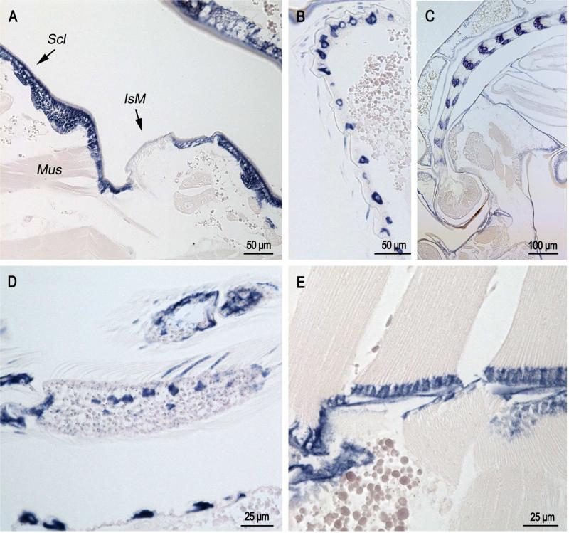
Fig. 3.
In situ hybridization of AgamCPCFC1 on sections of pupae less than 1 hour after pupation. A. Section of abdomen showing epidermal hybridization in sclerites (Scl) and only in intersegmental membrane (IsM) where muscles (Mus) are inserting into the cuticle. B. Lateral surface of pupal abdomen with setae-forming cells. C. Developing antenna in pupa. Structure was recognized because it is similar to that shown in Fig. 76a of Harbach and Knight (1980). D. Limb with developing scales showing hybridization. E. Muscle insertion zone with strong hybridization. (B,D 3’ probe; A,C,E coding region probe.)

