Figure 1.
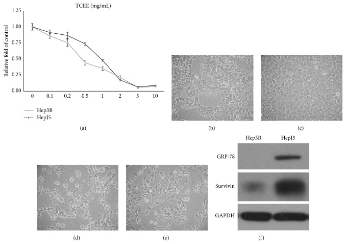
Cell growth inhibition of TCEE on human hepatocellular carcinoma cells, Hep3B and HepJ5. (a) Hep3B (gray line) and HepJ5 (black line) cells were treated with 0 to 10 mg/mL TCEE for 48 hr, and the cell viability was determined by MTT assay. IC50 of TCEE is 0.48 mg/mL on Hep3B cells and 0.91 mg/mL on HepJ5 cells, respectively. Experiments were repeated in triplicate and presented data were mean plus standard deviation. ((b) to (e)) Morphological observation on Hep3B and HepJ5 cells treated with 0 mg/mL TCEE ((b) and (c) Hep3B and HepJ5, resp.) or 0.5 to 1.0 mg/mL TCEE for 48 hr ((d) and (e) Hep3B and HepJ5, resp.). TCEE treated cells demonstrated apoptotic-like morphological changes such as cell shrinkage and cell blebbing compared with cells treated with normal culture medium. Magnification = 100x. (f) Expressions of survivin and GRP-78 on Hep3B and HepJ5 cells were determined by western blotting analysis. HepJ5 cells demonstrated higher expression of both survivin and GRP-78 than Hep3B cells. GAPDH served as the internal control.

