
Figure 4. Analysis of globin gene and protein expressions the corrected erythrocytes, and their function.
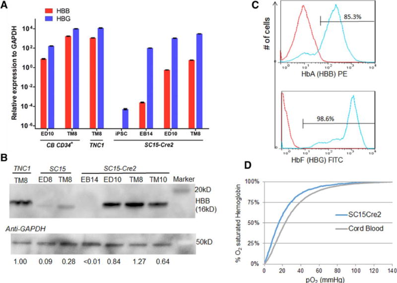
(A) Quantitative RT-PCR analysis of HBB and HBG gene expression in iPSCs, after hematopoietic differentiation (EB14), erythroid differentiation (ED10) and terminal maturation (TM8) from SC15-Cre2 and parental TNC1 iPSCs and a control of cord blood (CB) CD34+ cells. (B) Western blot analysis of adult hemoglobin beta (HBB) protein level during the differentiation of iPSC clones before and after correction and excision. The relative levels of full-length HBB proteins normalized to the GAPDH control are also indicated. (C) FACS analysis of intracellular adult and fetal hemoglobin protein expression of maturated erythrocytes generated from SC15-Cre2 iPSC clone. (D) Oxygen affinity curves of erythrocytes from SC15-Cre2 after terminal maturation and a cord blood control as measured by a Hemox Analyzer.