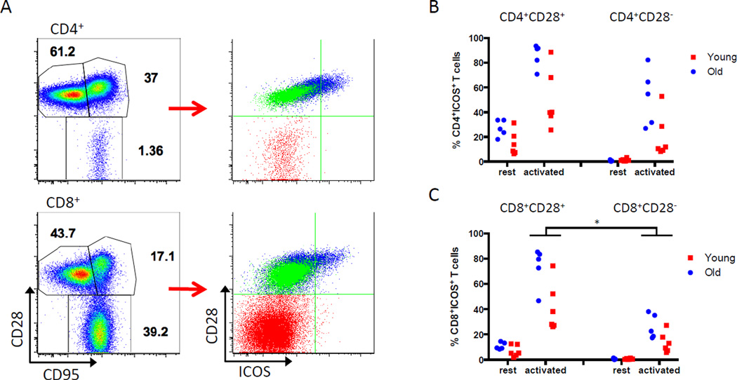
Figure 2. Expression of ICOS in resting and activated memory T cell subsets.
PBMCs from 11 healthy, young (n=6) and older (n=5) rhesus monkeys were analyzed for ICOS expression by flow cytometry. (A) Memory subsets were defined using CD28 and CD95 expression (left) to derive naïve T cells (CD28+CD95lo; TN); central memory T cells (CD28+CD95hi; TCM); and effector memory T cells (CD28−CD95hi; TEM). Memory subsets were re-defined by CD28 and ICOS expression and overlaid (right). Resting TCM cells (blue overlay) express ICOS, while TN cells (green) and TEM cells (red) do not express ICOS at rest. Representative data from a young monkey is shown. (B, C) PBMCs were stimulated for 24 hours with PMA/ionomycin. CD4+ (B) and CD8+ (C) CD28+ and CD28− subsets were analyzed for ICOS expression. Data is expressed as % ICOS+ T cells in CD4+CD28+, CD4+CD28−, CD8+CD28+ or CD8+CD28− subsets. Activated CD8+CD28− T cells expressed significantly less ICOS than CD8+CD28+ T cells (* p=0.0001).

