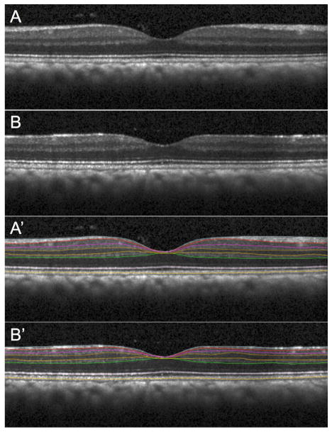
Figure 5. Macular retinal layer thickness parameters obtained from SDOCT radial scans under manometric IOP control.
Individual example of an NHP EG eye imaged at baseline (A, A’) and at its final time point (B, B’) just prior to sacrifice when glaucomatous damage was severe (as determined by complete orbital optic nerve axon counts). One of 48 B-scans (vertical orientation) from a radial pattern centered on the fovea is shown without segmentation in A and B, with layer segmentations in A’ and B’. This example is representative of the pattern of results observed across N=21 NHP in this study: there was clear, consistent loss in EG eyes of macular nerve fiber layer, ganglion cell layer and inner plexiform layer thickness, along with a small but significant increase in this thickness of both the outer plexiform layer and a layer defined as the outer nuclear (photoreceptor cell) layer plus inner segments and Henle’s fibers, with no change in outer segment length (defined as being from the reflectivity band at the inner segment-outer segment junction to the band at the cone outer segment tips, “COST line”). Macular inner retinal layer thickness losses were strongly correlated with peripapillary RNFL thickness in EG eyes but the subtle increase of outer retinal layer thickness was only weakly correlated with loss peripapillary RNFL thickness. Macular inner retinal layer losses were correlated with loss of function specific to retinal ganglion cells derived from multifocal electroretinography, while function of distal retinal elements was unchanged relative to fellow control eyes and un-related to peripapillary RNFL thickness loss. From Wilsey et al., Invest Ophthalmol Vis Sci 2015;56: E-Abstract 638.

