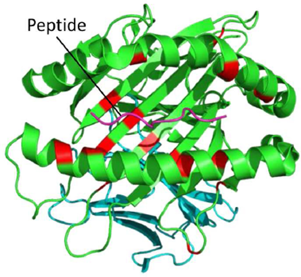
Figure 1. The MHC class I molecule, HLA-B, showing BD-associated amino acid positions.
A 3D model of the HLA-B molecule drawn by PyMol using 1E27, protein data of HLA-B*51:01 from the Protein Data Bank. The pink line shows a peptide antigen in the antigen-binding groove of the HLA-B molecule. Red indicates amino acids with genome-wide significant association (P < 5 × 10⁻⁸) with BD from [43].

