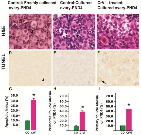
Fig. 3.
Effects of CrVI on follicle atresia in cultured whole fetal ovaries on culture day 12. Fetal ovaries from embryonic day (E) 13.5 were cultured for 11 days in vitro, and treated with CrVI (0.1 ppm) from CD2 to CD8, and harvested on CD12 (which recapitulated PND4). Ovaries from culture were harvested and fixed in 4% buffered paraformaldehyde and processed for histology (A-C) or in situ TUNEL apoptotic assay (D-F). The apoptosis index (AI) was calculated as the average percentage of TUNEL-positive oocytes from 10 ovaries. Average number of TUNEL-positive cells in control group was considered as 10%. Number of atretic primordial or primary follicles were counted in serial sections and expressed in percentage. *: control vs CrVI, p<0.05. The width of field for each image is 220 or 350 µm. Arrow heads point out healthy follicles. Arrows point out atretic follicles. Representative images and histograms are shown.

