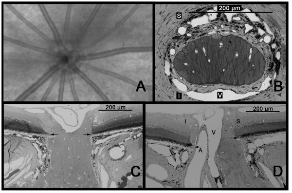
Figure 1.
Anatomy of the rat ONH. A. Anterior view of the rat fundus as seen by Optical Coherence Tomography (OCT). The ONH is dominated by spoke-like retinal arteries and veins that all but obscure the actual neural portion of the ONH. B. Cross-sectional view of the ONH at the level of the sclera. The nerve is horizontally oval and densely surrounded by vessels, including the central retinal vein (V), which is located inferiorly. The central retinal artery (not seen) lies inferior to the vein. C. Horizontal longitudinal section shows the optic nerve contacting the edge of Bruch’s membrane at either extreme. D. A vertical longitudinal section shows that only the superior aspect of the nerve is in contact with the edge of Bruch’s membrane, while inferiorly, the ONH is separated from Bruch’s by the central retinal artery as well as the vein. Arrows = Bruch’s membrane; S = superior; I = inferior; A = central retinal artery; V = central retinal vein. (A. Courtesy of R. Wang and Z. Zhi)

