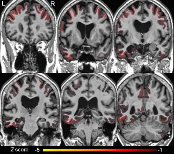
Figure 2.
MRI atrophy patterns using differential diagnosis based on structural abnormality in neurodegeneration (differential-STAND).
There is atrophy in bilateral mid and superior frontal regions, bilateral precentral regions (worse on the left), bilateral precuneus, and postcentral regions. The left hippocampus, fusiform and mid temporal regions also showed significant atrophy.
Abbreviations (z score refers to the number of standard deviations): L, left; R, right (note for this analysis the normal MRI left-right orientation is reversed; the left brain is on the left of the figure and the right brain is on the right of the figure for each panel).

