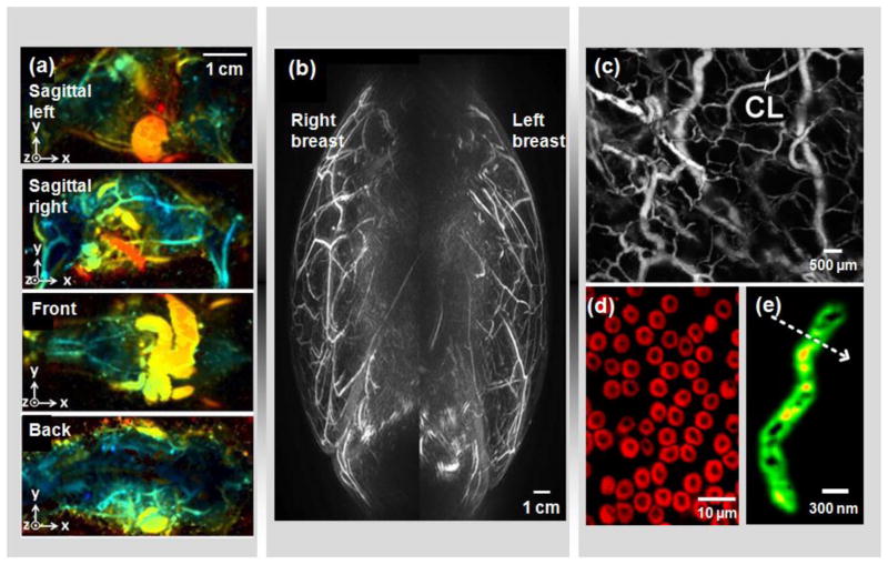
Figure 2. Multi-scale PAT.
(a) Whole-body PAT of a mouse in vivo [43]. Acquired at 1064 nm, the maximum amplitude projection (MAP) images were extracted from sagittal, front and back views. The depths are color encoded from blue (shallow) to red (deep). (b) Medial-lateral MAP image of breasts of a healthy volunteer [12]. A semi-spherical transducer array was used with rotational scanning. (c) Optical-resolution photoacoustic microscopy (OR-PAM) of mouse ear vasculature, where single capillaries (CL) can be clearly resolved [64]. (d) Sub-wavelength OR-PAM of single red blood cells [75]. A lateral resolution of ~220 nm was achieved. (e) Photoacoustic nanoscopy of a mitochondrion in a fibroblast cell [208]. A lateral resolution of ~80 nm was achieved by using the optical absorption saturation effect. Images were adapted with permission from references [12, 43, 64, 75, 208].

