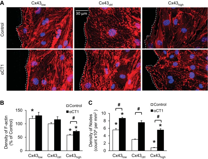
Fig. 5.
Cx43 modulates F-actin architecture through ZO-1 binding. A: representative images of cells at wound edges, delineated with dashed lines. Wound space is to the left. The density of cytoskeletal F-actin staining with phalloidin (red) was inversely proportional to the level of Cx43. Disrupting the Cx43/ZO-1 complex with αCT1 caused a distinct change in F-actin architecture characterized by a high density of nodes in all conditions. B and C: quantification of the density of F-actin (B) and F-actin nodes (C) in cells at wound edges using ImageJ. *P < 0.05 vs. Cx43ctrl; #P < 0.05 for Control vs. αCT1; n = 6 per mean.

