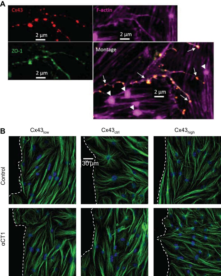
Fig. 6.
Microtubule architecture is not grossly altered by Cx43/ZO-1 uncoupling. A: Cx43 (red) and zonula occludens-1 (ZO-1; green) are colocalized (arrows) along continuous strands of F-actin (purple). F-actin nodes (arrowheads) occur only in the absence of Cx43/ZO-1 colocalization. B: representative images of microtubules (β-tubulin) in cells at wound edges with varying levels of Cx43; wound space is to the left, delineated with dashed lines. The architecture of cytoskeletal microtubule labeling with an antibody to tubulin (green) under control conditions and following disruption of the Cx43/ZO-1 complex with αCT1. Nuclei are shown in blue.

