Fig. 4.
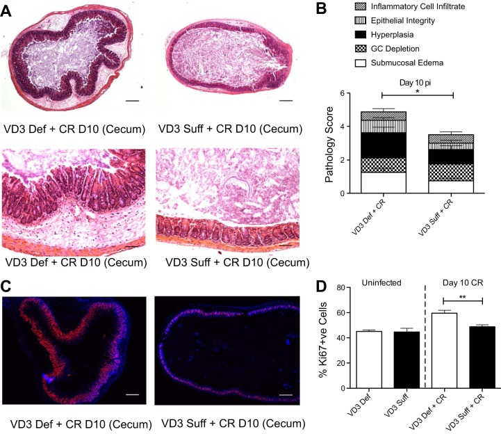
Vitamin D3-deficient mice suffer worsened histological damage with increased cell proliferation in the ceca at day 10 pi with C. rodentium. Weanling (3-wk-old) female C57Bl/6 mice were fed vitamin D3-deficient (0 IU) or vitamin D3-sufficient (1,000 IU) diets for 5 wk and then orally infected with C. rodentium. A: representative image of cross section of the cecum at day 10 pi. Original magnification = ×50 for top and ×200 for bottom. Scale bar = 200 μm. B: cecum was assessed for histological damage by scoring system for C. rodentium described in materials and methods. GC, goblet cell. Results are representative of 3 independent experiments; n = 8 per group; *P < 0.05 by Mann-Whitney test. C: representative images of formalin fixed cross section of cecum at day 10 pi. Immunofluorescence stained: blue, DAPI; red, Ki67. Original magnification = ×50; scale bar = 200 μm. D: cecum was assessed for number of Ki67+ve cells in the lumen (percent of +ve Ki67 per +ve DAPI per area measured). Results are representative of 3 independent experiments; n = 4 per group (uninfected) and n = 11 per group at day 10 pi; **P < 0.01 by Mann-Whitney test.

