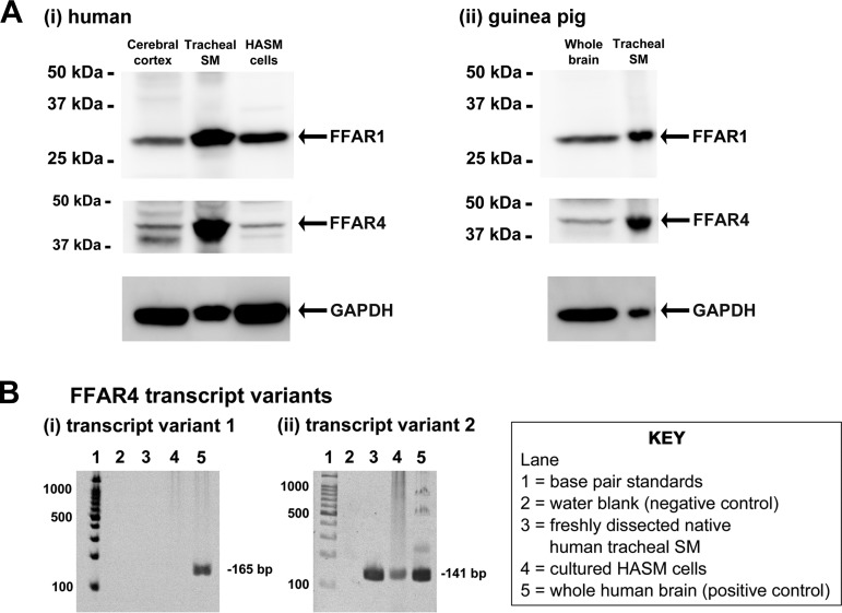
Fig. 1.
A: representative gel images of immunoblot analyses using antibodies against the free fatty acid receptor 1 (FFAR1) and FFAR4 using total protein prepared from human (i) and guinea pig (ii) tissues: human brain cerebral cortex (50 μg), freshly dissected native human tracheal airway smooth muscle (SM; 100 μg), primary cultured human airway smooth muscle (HASM) cells (100 μg), guinea pig whole brain (150 μg), and freshly dissected native guinea pig tracheal SM (100 μg). Reprobing of blots for GAPDH was performed to demonstrate relative lane loading. B: representative gel images of RT-PCR analyses of total RNA using primers specific for human FFAR4 transcript variant 1 (i) and variant 2 (ii). Total RNA extracted from freshly dissected human tracheal SM or cultured HASM cells was analyzed. Lane 1, base pair standards; lane 2, negative control water blank; lane 3, total RNA from freshly dissected native human tracheal SM; lane 4, total RNA from primary cultured HASM cells; lane 5, total RNA from whole human brain (positive control).

