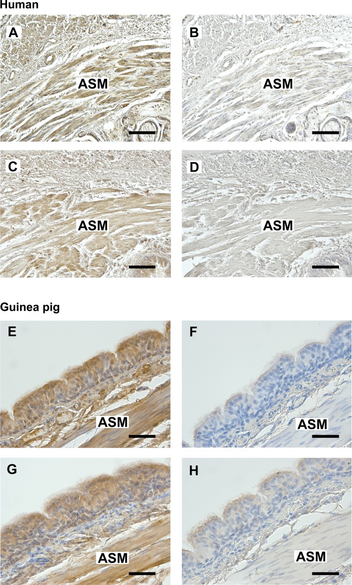
Fig. 2.
A and C: representative photomicrographs of immunohistochemical staining of FFAR1 (A) or FFAR4 (C) in paraformaldehyde-glutaraldehyde-fixed human trachea. E and G: representative photomicrographs of immunohistochemical staining of FFAR1 (E) or FFAR4 (G) in paraformaldehyde-fixed guinea pig trachea. B and D: anti-rabbit IgG isotype negative control in serial section of human trachea. F and H: anti-rabbit IgG isotype negative control in serial section of guinea pig trachea. All sections were counterstained with hematoxylin. Calibration bars: A–D, 100 μm; E–H, 50 μm. ASM, airway smooth muscle. Images are representative of at least 3 independent immunohistochemical analyses from both human and guinea pig trachea.

