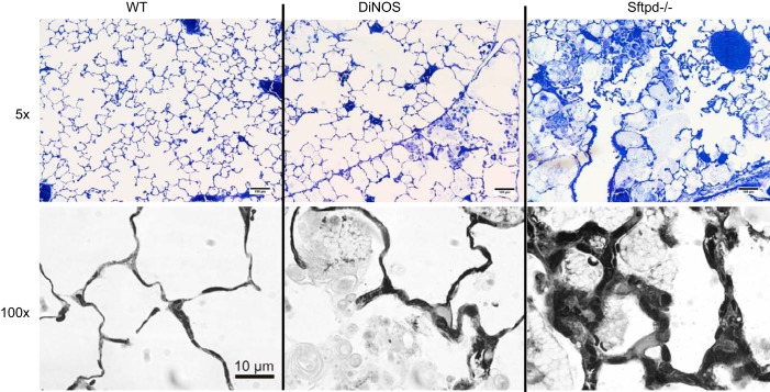
Fig. 3.
Inflammatory cells, alveolar lipoproteinosis, and focal septal wall thickening. Representative light microscopic images of n = 4–5 animals/group. Normal lung architecture can be seen in WT mice. In DiNOS mice, slight enlargement of distal airspace filled with alveolar macrophages and intra-alveolar surfactant material are typical findings. In Sftpd−/− mice, these alterations are much more prominent; ×100 oil immersion light microscopic images show pathological alterations in Sftpd−/− and DiNOS. Septal walls are focally thickened in close proximity with foamy appearing alveolar macrophages in Sftpd−/− mice. Although enlarged alveolar macrophages are present in DiNOS, septal walls are much thinner compared with Sftpd−/− mice.

