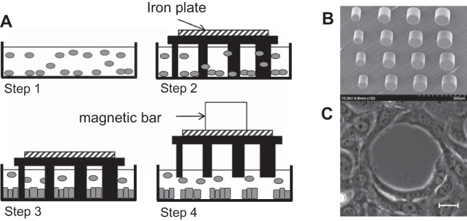
Fig. 1.

Gap creation with polydimethylsiloxane (PDMS) micropillars. A: experimental procedure for undamaged gap creation. Cells are seeded onto a collagen-coated glass surface (step 1). Then a PDMS stencil coated with a Pluronic block copolymer is placed on the dish (step 2) and incubated for 1 day to enable cells to attach to the glass surrounding the pillars (step 3). Finally, stencil detachment with a magnetic bar creates undamaged gaps of different sizes (step 4). B: scanning electron microscopy of a stencil of micropillars. C: gap pattern on a bovine aortic endothelial cell (BAEC) monolayer. Scale bar, 10 μm.
