Fig. 3.
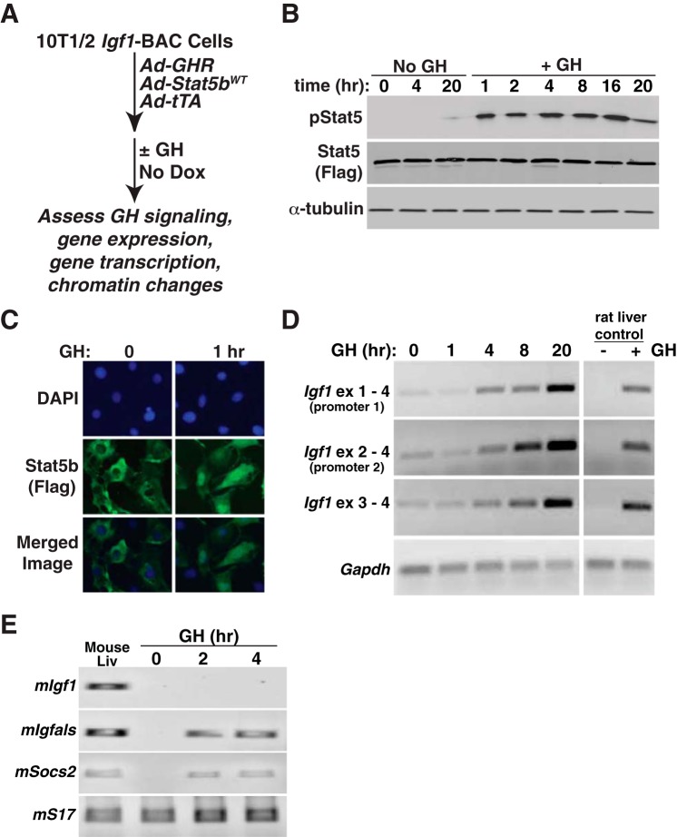
Rapid induction of Igf1 gene expression by GH in a BAC-containing cell line. A: experimental plan outlining steps to test Igf1 gene regulation in response to GH and Stat5b in 10T1/2 Igf1-BAC clone 11. B: time course of appearance of tyrosine phosphorylated Stat5b (pStat5), total Stat5b (using antibody to Flag), and α-tubulin, as assessed by immunoblotting after addition of vehicle (No GH) or recombinant rat GH [40 nM] to clone 11 cells. C: GH treatment causes the nuclear accumulation of Stat5b in the nucleus of 10T1/2 Igf1-BAC clone 11, as detected by immunocytochemistry. DAPI was used to stain nuclei. D: time course of accumulation of Igf1 and Gapdh mRNAs, as assessed by RT-PCR using RNA isolated at different times after GH addition to cells. Liver RNA from GH-deficient rats treated acutely with vehicle or a single injection of recombinant rat GH [1.5 mg/kg] serves as both negative and positive controls for GH-responsiveness. E: time course of appearance of mouse Igf1, Igfals, Socs2, and S17 mRNAs, as assessed by RT-PCR using RNA isolated at different times after GH addition to cells. Liver RNA from intact adult male mice serves as a positive control.

