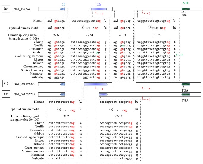
Figure 4.
MYEOV splicing junctions. Illustrated are three MYEOV alternatively spliced variants (a–c), as described in Figure 1. Shown are the canonical 5′/3′ splice sites that delimit all the putative splicing junctions of MYEOV, as annotated in the Gene database from NCBI. The splice sites are juxtaposed to the optimal core splicing signal motifs; corresponding splicing signal strength values are also shown. Upper-case nucleotides, delimited by brackets, versus lower-case nucleotides correspond to exonic versus intronic content, respectively. Nucleotides exerting the strongest influence on the signal strength appear in red font. Aligned below the human splicing signals appear corresponding nucleotide sequences from MYEOV syntenic region in 11 primates. Shown in green font are nucleotides that deviate from the evolutionary trend, requiring further validation. The double-headed green arrow points to the precious canonical acceptor that arose in hominoids, allowing for MYEOV long-ORF to occur. Alignment gaps correspond to indels located between the aligned blocks in the aligning species.

