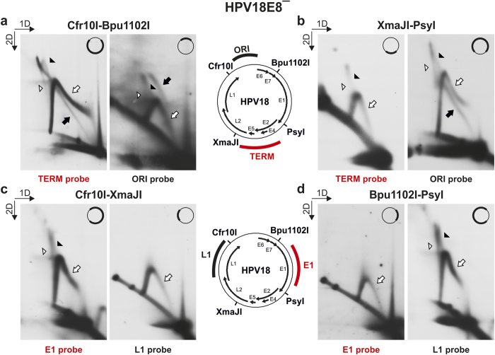
Figure 5. 2D N/N AGE analysis of RIs arising from subgenomic fragments of HPV18E8ˉ episomes extracted from U2OS cells 3 days post-transfection.
The positions of the recognition sites of used restriction endonucleases Cfr10I, Bpu1102I, PsyI and XmaJI are marked in both schemes. The HPV18 genomic areas specific for the ORI and TERM hybridization probes used for the analysis of Cfr10I-Bpu1102I and XmaJI-PsyI fragments (a,b) are marked in the upper scheme; the areas specific for the E1 and L1 probes used for the analysis of Cfr10I-XmaJI and Bpu1102I-PsyI fragments (c,d) are marked in the lower scheme. A simplified version of the schemes is in the upper right corner of each figure, with the analyzed fragment marked in bold. Black arrowheads, putative late theta RIs; white arrowheads, X-shaped molecules; black arrows, theta RIs; white arrows, intermediates of the second replication mechanism. The direction of the gel electrophoresis in the first (1D) and second (2D) dimension is indicated in the top left corners of the panels. (a) 2D N/N AGE of the Cfr10I-Bpu1102I fragments. (b) 2D N/N AGE of the XmaJI-PsyI fragments. (c) 2D N/N AGE of the Cfr10I-XmaJI fragments. (d) 2D N/N AGE of the Bpu1102I-PsyI fragments.

