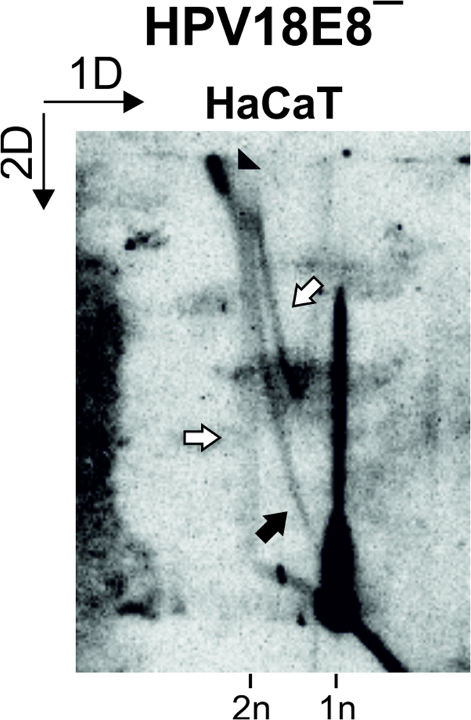
Figure 7. Analysis of RIs arising during the initial amplification replication of HPV18E8ˉ episomes in HaCaT cells.

LMW DNA was extracted from HaCaT cells 5 days post-transfection, digested with BglI and analyzed via 2D N/N AGE. Black arrowhead, putative late theta RIs; black arrow, theta RIs; white arrows, intermediates of the second replication mechanism. 1n, monomeric (8-kbp) linear molecules; 2n, dimeric (16-kbp) linear molecules. The direction of the gel electrophoresis in the first (1D) and second (2D) dimension is indicated in the top left corner.
