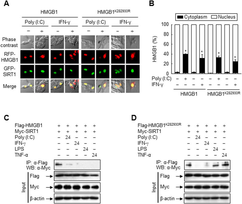
Figure 6. Localizations of HMGB1 and SIRT1 in CHO cells treated with Poly (I:C) or IFN-γ.
(A,B) CHO cells co-transfected with GFP-SIRT1 and RFP-HMGB1 or RFP-HMGB1K282930R for 48 h were incubated with Poly (I:C) (50 μg/ml) or IFN-γ (40 ng/ml). Following incubation for 24 h, the fluorescence of each fusion protein was visualized by confocal microscopy (A) and quantified (B). The bar indicates 30 μm. The co-localization of HMGB1 and SIRT1 is indicated by the presence of yellow in the merge images. Results are expressed as the means ± standard error (n = 3). *p < 0.01 compared with the untreated group. (C,D) HEK293T cells co-transfected with Myc-SIRT1 and wild-type or mutant Flag-HMGB1 for 48 h were incubated with Poly (I:C) (50 μg/ml), IFN-γ (40 ng/ml), LPS (100 ng/ml), or TNF-α (20 ng/ml) for 24 h, and then whole-cell lysates were prepared and immunoprecipitated with an anti-Flag antibody. The immunoprecipitates and total lysates (input) were subjected to immunoblot analysis with anti-Flag, anti-Myc, and anti-β-actin antibodies to detect HMGB1, SIRT1, and β-actin, respectively. Two percent of whole-cell lysates were used as the input.

