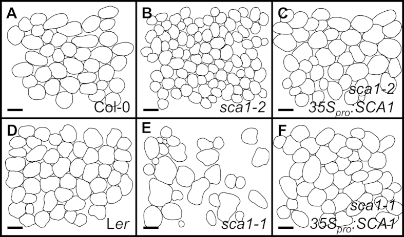
Figure 3. Representative diagrams of the sub-epidermal layer of palisade mesophyll cells from a first leaf of the (A) Col-0 and (D) Ler wild types, the (B) sca1-2, and (E) sca1-1 mutants, and the 35Spro:SCA1 transgenic line in the (C) sca1-2 and (F) sca1-1 genetic backgrounds, respectively.
Diagrams were drawn from differential interference contrast images taken from cleared leaves. Scale bars indicate 50 μm.

