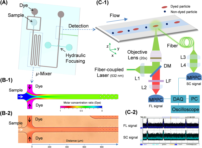
Figure 1. Micro-optofluidic platform for real-time, continuous detection of airborne microorganisms.
(A) A schematic diagram of the structure of the microchip flow cytometer. It consists of a sample inlet, a micro-mixer, and a detection component in a single layer of PDMS, and fabricated using standard soft lithographic methods. (B-1) Simulated mixing conditions between the sample and the dye in the micro-mixer. The diffusivity coefficient of the dye in water was 10−9 m2/s. The microfluidic flow rates of both the sample and dye suspension were 12.5 μL/h. The simulations were carried out using CFD-ACE+. (B-2) Optical micrograph of the results corresponding to the simulation shown in B-1. (C-1) Real-time detection of target particles in the core flow in the detection zone, and the optical setup used for continuous particle detection and signal quantification. The target particles in the core of the sample flow were exposed to a fiber-coupled 532-nm laser. The emitted fluorescent (FL) light was collected and brought to the MPPC photo sensor via an optical fiber. A multi-mode fiber was inserted into the PDMS chip to guide the particle scattering (SC) light to the MPPC photo sensor. L1: fiber collimator, f = 7.5 mm; L2: plano-convex lens, f = 50 mm; L3: fiber collimator, f = 18.1 mm; L4: plano-convex lens, f = 25.4 mm; DM: dichroic mirror (552 nm); LF: long-pass filter (560 nm). (C-2) Optical signals for FC and SC monitored using an oscilloscope connected to the MPPC.

