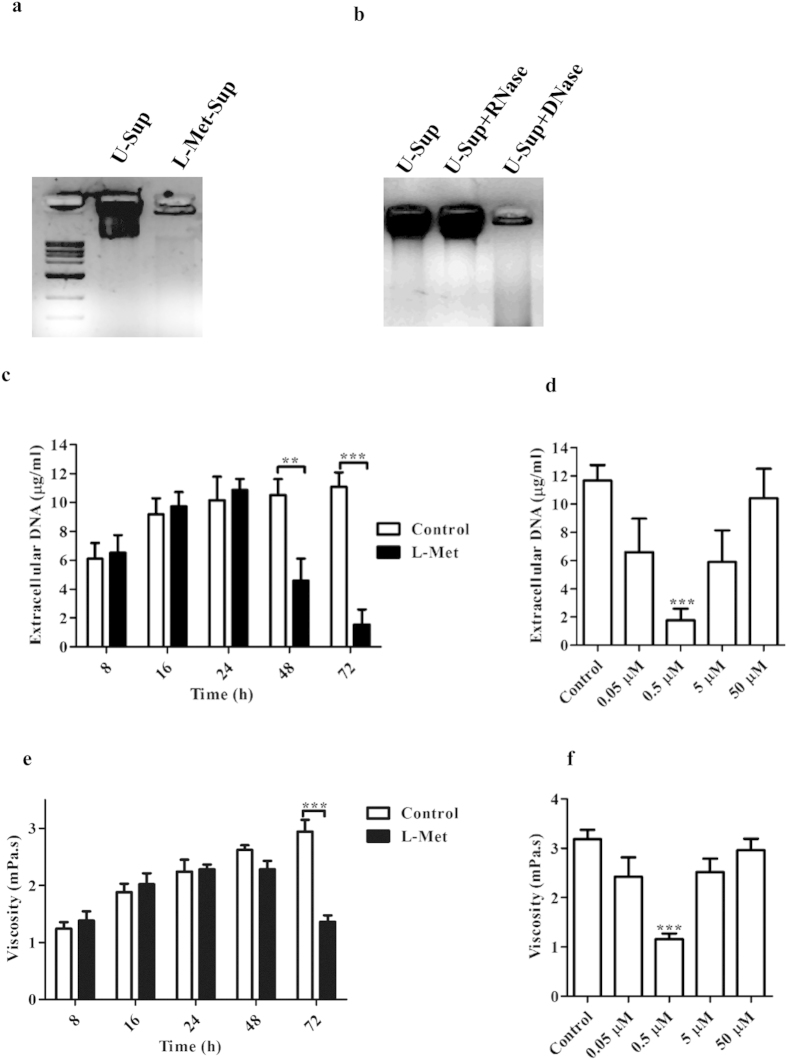
Figure 2. L-Methionine (L-Met) degrades extracellular DNA.
72 h old PA biofilm culture media was centrifuged at 2000 g for 5 min and the supernatant of (a) Untreated culture (U-Sup) & (b) L-Met treated culture (L-Met-Sup) were loaded in the agarose gel and visualized. (b) U-Sup was treated with DNase and RNase (Fermentas) for 1 h at 37 °C and then loaded in the agarose gel and visualized. Extracellular DNA was precipitated and DNA concentration was determined by spectrofluorimetrically from U-Sup and L-Met-Sup (c) with different duration of incubation with L-Met (0.5 μM) or (d) with different concentrations of L-Met and incubated for 72 h. Viscosity of U-Sup and L-Met-Sup (e) with different duration of incubation with L-Met (0.5 μM) or (f) with different concentrations of L-Met and incubated for 72 h was determined using Rheometer. (D & F - 0.5 μM was compared with control). Statistical significance was calculated using One-way ANOVA. Asterisks indicate statistical significance as follows: **(p < 0.005), ***(p < 0.001).

