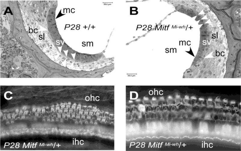
Figure 3.

Morphology of +/+ and MitfMi-wh/+ stria vascularis and cochlear hair cells. Examples from P28 MitfMi-wh/+ mouse (B) and +/+ littermate (A) are shown. (A) Stria vascularis of P28 wild-type mouse. Arrowheads point to the condensed marginal cell (mc) nuclei and basal cell (bc) nuclei of the stria vascularis, with grouped arrowheads defining the boundary between the stria vascularis (sv) and the spiral ligament (sl). The boundaries between cell layers in the stria vascularis are obscured by faint interdigitations. (B) Stria vascularis of P28 MitfMi‐wh/+ mouse. In this panel, the grouped arrowheads delineate the sharp boundary present between the marginal and basal cell layers. The marginal cell (mc) nuclei are rounded rather than condensed. (C) Apex of P28 MitfMi‐wh/+ cochlea. Whole-mount phalloidin staining reveals no inner hair cell (IHC) loss, with sporadic loss of outer hair cells (OHCs). (D) Whole-mount phalloidin staining of base of P28 MitfMi‐wh/+ cochlea shows similar OHC loss with preservation of IHC layer. Other symbols: sm, scala media.
