
Figure 5.
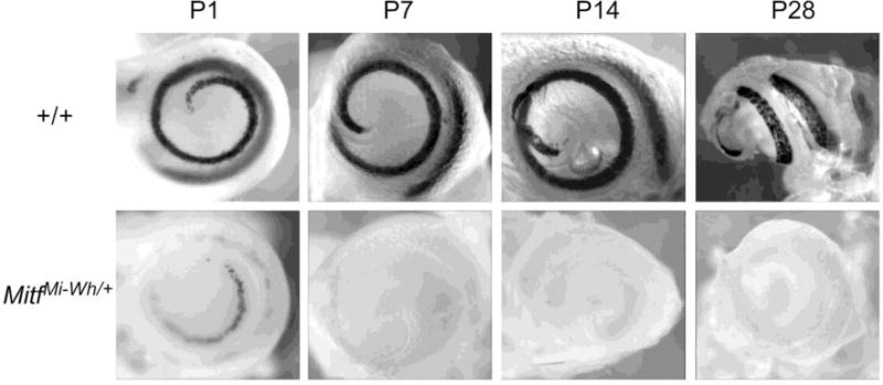
Stria vascularis melanocytes in neonatal +/+ and MitfMi-wh/+ mice. +/+ and MitfMi‐wh/Mi-wh mice were intercrossed with Dct-lacZ transgenic mice to permit visualization of cochlear melanocytes after staining for β-galactosidase activity. Wild-type cochlear melanocytes are present in a continuous, 1 3/4 turn configuration in whole-mount P1, P7, P14, and P28 cochleas (top row). MitfMi-wh/+ cochlear melanocytes (bottom row) are more sparse and distributed intermittently along the stria vascularis at P1, yet still present in a similar distribution as in the wild-type cochlea. In most P7cochleas and in older (P14 and P28) cochleas, MitfMi-wh/+ melanocytes (left) are no longer visible.