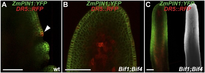
Fig. S2.

Confocal analysis of ZmPIN1a:YFP and DR5rev::RFP transgenes. (A) Wild-type tassel, showing the peripheral zone of the IM with an emerging primordium (arrowhead). (B and C) +/Bif1;+/Bif4 tassels. In B is a close-up view of the IM. (C, Right) Brightfield image of the confocal sample. (Scale bars, 100 μm.)
