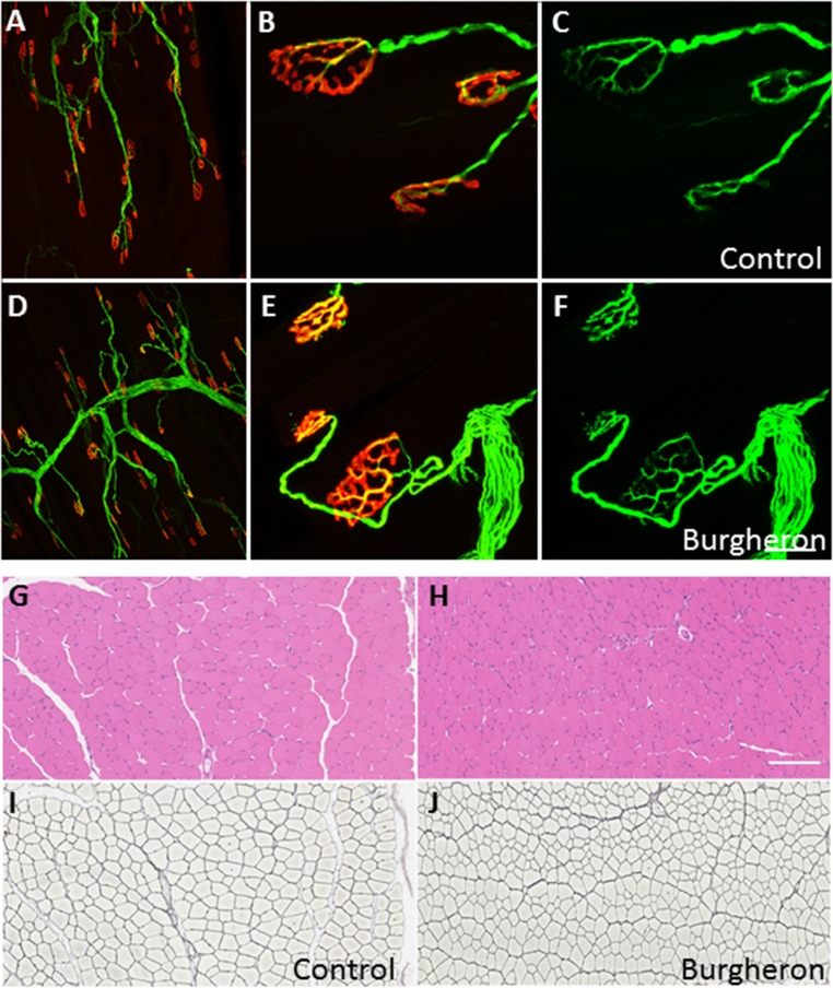
Fig. S2.
Normal innervation of the NMJs and normal muscle histology in the Burgheron mutants. Control (A–C) and mutant (D–F) endplates with immunostaining for antineurofilament and antisynaptic vesicles (green) and alpha-bungarotoxin staining of AChRs (red). Low (A and D) and high (B and E) magnifications show a normal apposition of the nerve terminal with the postsynaptic receptors with no sign of denervation. When singled out in the green channel (C and F), the nerve terminal displays no sign of neurofilament accumulation, retraction, or sprouting in the mutants. Shown are control (G and I) and mutant (H and J) TA muscle cross-sections stained with hematoxylin/eosin (G and H) and for reticulin (Gordon Sweet staining; I and J). The reticulin stain outlines individual muscle fibers. No sign of severe muscle atrophy or necrosis was observed, although muscle fibers were on average smaller in the mutants. All mice were imaged at P35–P40. [Scale bar, (A and D) 600 μm, (B, C, E, and F) 20 μm, and (G–J) 250 μm.]

