Fig. 9.
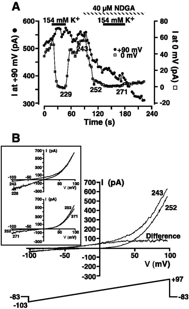
Isolation of the MIP current in low extracellular K+ solution by NDGA inhibition. A: cell suspended in 149 Na+/5 mM K+ solution (solution 9) was whole cell patch-clamped with a Mg2+-free KCl-based internal solution (solution 3). The 200-ms voltage-ramp protocol administered every 3 s is shown in B. Magnitude of the outward current at +90 and 0 mV was extracted from ramp data and plotted as a function of time. Where indicated, extracellular solution was changed to 154 mM K+ (solution 10), and 40 μM NDGA was applied. B: I-V relationships in 149 Na+/5 mM K+ solution (solution 9) before (episode 243) and shortly after (episode 252) application of 40 μM NDGA. I-V relationship labeled “Difference” was obtained by subtraction of the I-V relationship after NDGA addition (episode 252) from that before NDGA addition (episode 243) and is the I-V relationship for the MIP conductance in the absence of TRPM7 contamination. Inset: I-V relationships obtained in the presence (episodes 229 and 271) and absence (episodes 243 and 252) of high extracellular K+ solution before and during NDGA application as denoted in A. Voltage-ramp protocol is shown in main panel.

