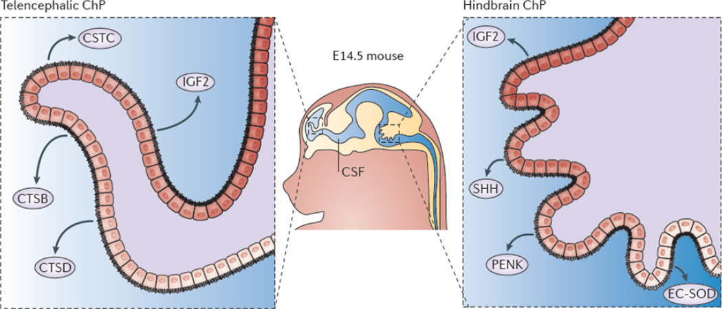
Figure 4. Telencephalic and hindbrain choroid plexi are transcriptionally and functionally distinct tissues.
Gene expression profiling of mouse and primate telencephalic choroid plexus (ChP) and hindbrain ChP reveal that, despite being morphologically similar, these tissues are transcriptionally heterogeneous35. The ChPs maintain distinct positional identities reminiscent of their axial tissues of origin. The telencephalic ChP has higher expression levels of Emx2Otx1, and Six3, which are markers of the telencephalon, whereas the hindbrain ChP has higher expression levels of Hoxa2En2, and Meis1, genes critical for patterning the hindbrain. Further analysis of the ChP secretome reveals that these tissues are functionally distinct in their expression and secretion of signalling factors into the cerebrospinal fluid (CSF)35. In particular, the telencephalic ChP secretes more cystatin C (CSTC), cathepsin B (CTSB) and cathepsin D (CTSD), whereas the hindbrain ChP secretes more Sonic hedgehog (SHH), proenkephalin (PENK) and extracellular superoxide-dismutase (EC-SOD). Protein expression domains have been observed within each ChP (for example, EC-SOD35 and SHH53 protein domains), which is suggestive of protein gradients within each ventricle. These findings indicate that the molecular heterogeneity of the telencephalic versus the hindbrain ChP contributes to establishing a regionalized CSF and support a model of protein gradients throughout the ventricular system.

