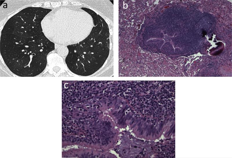
Fig. 10.
Small airway disease, follicular bronchiolitis. 40-year-old female with rheumatoid arthritis whose pulmonary function test showed marked obstructive pattern. (a) CT. Mild bronchial wall thickening and mosaic attenuation in both lungs suggesting air trapping. (b and c), Transbronchial biopsy. Dense peribronchial lymphoid infiltrate with follicular hyperplasia, causing bronchiolar luminal narrowing and obliteration.
Teaching point: Imaging findings in symptomatic patients with small airways disease can be very subtle. Tailoring of CT protocols and use of paired inspiratory and expiratory imaging is helpful to accentuate air trapping.

