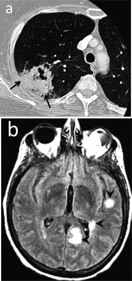
Fig. 15.
Angioinvasive aspergillosis. 52-year-old female with history of rheumatoid arthritis (RA) with fever, shortness of breath and seizure. (a) CT. Consolidation with cavitation in the right upper lobe represents invasive aspergillosis (arrows). Aspergillus was isolated from bronchoalveolar lavage. (b) Contrast-enhanced T1-weighted MR image of the brain. Multiple enhancing cerebral foci (arrowheads) were presumed to represent angioinvasive aspergillosis.
Teaching point: Opportunistic infection secondary to immunosuppression is not uncommon among RA patients.

