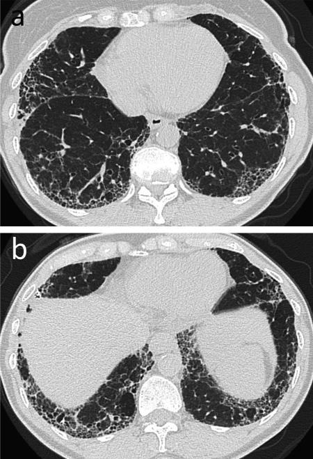
Fig. 6.
Usual interstitial pneumonia (UIP). 63-year-old female with long-standing history of rheumatoid arthritis. (a, b) CT. Bilateral lower lobe predominant subpleural reticular opacity and honeycombing. Biopsy confirmed the diagnosis of UIP. Teaching point: Multilicity of CT patterns may be seen; however, UIP is the most common pattern of interstitial lung disease in rheumatoid arthritis patients.

