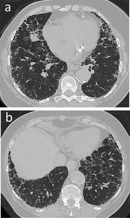
Fig. 7.
Nonspecific interstitial pneumonia (NSIP). 76-year-old male with longstanding history of rheumatoid arthritis with subcutaneous rheumatoid nodules. (a and b) CT. Moderate peripheral reticular opacities and groundglass opacities with minimal honeycombing predominantly in the lower lobes. Biopsy showed changes most consistent with fibrotic NSIP.
Teaching point: NSIP and UIP are common patterns of rheumatoid-associated inter-stitial lung disease. NSIP and UIP can have overlapping imaging features. In such cases, biopsy is required to make the diagnosis.

