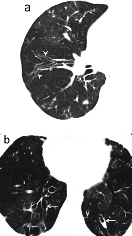
Fig. 9.
Large airway disease and progressive bronchiectasis. 58-year-old female with RA and severe bronchiectasis with superimposed MAI infection. (a) CT. Cylindrical bronchiectasis (arrowheads) with mosaic attenuation pattern. (b) CT obtained 6 years later. Progression of bronchiectasis, now varicose and cystic (arrows). Persistent areas of mosaic attenuation in both lungs suggest air trapping due to small airway involvement.
Teaching point: Rheumatoid arthritis is an important cause of non-cystic fibrosis bronchiectasis, which can predispose to subsequent infection.

