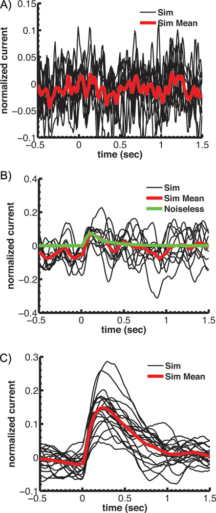
Figure 3.
Simulations of single-photon responses for a mouse rod with modified PDE kinetics or expression. Currents have been normalized to the circulating current in darkness. A: Simulations assuming a rate of spontaneous cGMP hydrolysis set equal to the diffusion-limited rate for light activated PDE. This high spontaneous PDE rate would give a βdark of approximately 55 s−1. B: Simulations of single-photon responses again assuming a rate of spontaneous cGMP hydrolysis set equal to the diffusion-limited rate for light activated PDE as in A but with a PDE density that is 13-fold lower than normal to reduce the amount of spontaneous PDE activations so that βdark is maintained at 4.1 s−1. C: Single-photon simulations with the PDE density reduced by a factor of 5 so as to be the same as in toad. All of A–C reprinted from Reingruber et al. [10] with permission of the authors, copyright 2013 National Academy of Sciences, U.S.A.

