
Fig. 1.
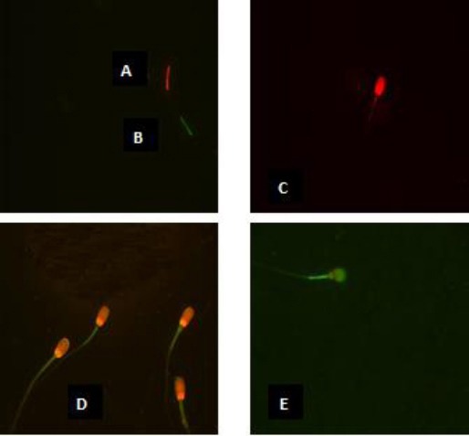
Spermatozoa viewed under phase contrast microscopy: spermatozoa alive and intact (colorless head) with high mitochondrial membrane potential (A); spermatozoa alive and intact (colorless head) with low mitochondrial membrane potential (B); dead spermatozoa (red head) with high mitochondrial membrane potential (C); dead spermatozoa (red head) with low mitochondrial membrane potential (D); reacted dead spermatozoa (red and green head) with low mitochondrial membrane potential (E).