
Figure 2.
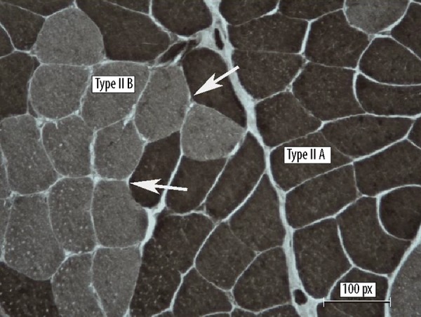
Muscle biopsy. Pattern of type grouping, reaction ATPasi pH 9,4 NADH-Tr (Hematoxylin-eosin (H&E), NADH (in black) show aspects of type grouping (enlargement ×100, Barr 1 μ). The arrows indicate clusters of new fibres. Fibres II A (black), Fibres II B (green).