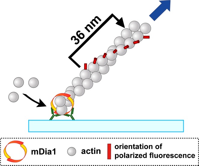
Figure 1.
Overview of visualization of rotational movement of an actin filament elongating from immobilized mDia1. GST-mDia1 FH1–FH2 was fixed in the protein aggregate composed of anti-GST antibodies and fluorescent secondary antibodies. The protein aggregate was adsorbed on the glass surface, and processive filament elongation was initiated by the addition of G-actin composed of TMR-actin (≈0.3%) and excess unlabeled actin. The vertically polarized fluorescence (FLV) and the horizontally polarized fluorescence (FLH) from TMR-actin in the filament were recorded after separated through a polarizing beam splitter.

