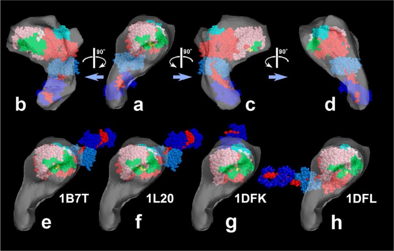
Figure 3.
Reconstructed shell to present 3-D envelope of the SH1–SH2 cross-linked myosin head [modified from Figure 7 of our paper26]. a)–d); Its tentative model was fit to the shell and viewed from four different angles. e)–h); Four known crystal structures of scallop-S1 whose catalytic domains are snugly placed in the shell of the novel structure. Note that the orientation of the lever-arm in the novel structure is directed quite differently from the others. The atomic models are, e) ADP-bound form, f) ADP-bound Lys705-Cys693 cross-linked form, g) nucleotide-free near-rigor form, h) ADP/Vi-bound form. See the legend to Figure 1 for color-coding of the subdomains.

