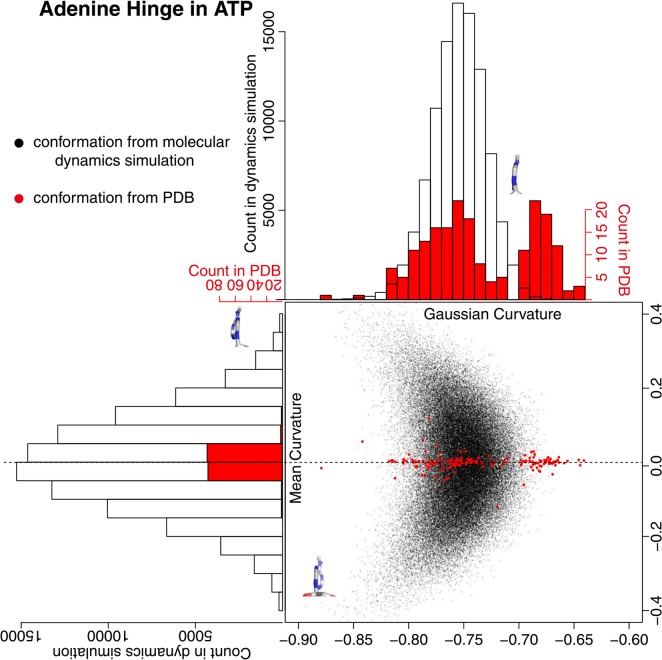
Figure 8.
Adenine hinge motion. The hinge motion is defined by the open-book movement in five-membered and six-membered rings in the adenine molecule. A pseudo-ring was defined to assess the openness of the hinge. See the method section for the detail. A black dot is obtained from the conformation of the molecular dynamics simulation, and a red dot is from PDB. The histogram in black clarifies the distribution of black dots and the one in red clarifies the distribution of red dots. The hinge conformations with minimum/maximum curvature values in the molecular dynamics simulation were drawn. A chemical bond at the bottom of each figure is a glycosyl bond and six-membered ring is located at the far side.

