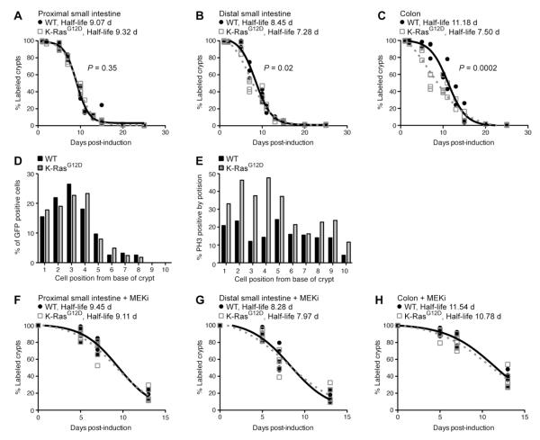
Figure 2.
K-RasG12D accelerates the loss of Egfp label in the intestinal epithelium. (A-C) Determination of Egfp half-life in animals expressing WT or mutant K-Ras. Animals were exposed to DOX (2 mg/ml) in the drinking water for 2 weeks and then sacrificed at defined time points. Immunohistochemistry for Egfp was used to identify crypts that included at least a single Egfp+ cell. Using these data, the Egfp half-life for control (black line) and K-Ras mutant animals (gray dotted line) was determined for the proximal small intestine (A), distal small intestine (B) and colon (C). A minimum of 3 animals were used for each experimental group at each time point, with approximately 50 crypts analyzed per animal. The Egfp half-life was significantly reduced by K-RasG12D in the distal small intestine (P = 0.0212) and colon (P = 0.0002), but not in the proximal small intestine (P=0.3510). (D) Position of Egfp label-retaining cells in WT and mutant animals. At time points near the half-life for each condition, immunohistochemistry for Egfp was used to determine the relative positions of Egfp+ cells within crypts that contain a single labeled cell. A minimum of 200 crypts was counted for each experimental group. Both experimental groups illustrated a similar distribution of Egfp positive cells within the crypt. (E) Distribution of mitotic cells in WT and mutant animals. The relative positions of phospho-Histone H3 (PH3) positive cells within crypts was determined by immunohistochemistry for PH3. A minimum of 200 cells at each position were counted for each experimental group. K-Ras mutant animals (gray line) displayed a higher percentage of PH3-positive mitotic cells than control animals (black line), particularly at cell position 1-5, were stem cells reside. (F-H) Determination of Egfp half-life after MEK inhibition in animals expressing WT or mutant K-Ras. Animals were treated daily with PD0325901 (MEKi) for either 5, 7, or 13 days following DOX pulse. The half-life for both control (black line) and K-Ras mutant animals (gray dotted line) was determined for the proximal small intestine (F), distal small intestine (G) and colon (H). 5 animals per time point were used for each experimental group. In the presence of MEKi, K-RasG12D does not affect label retention.

