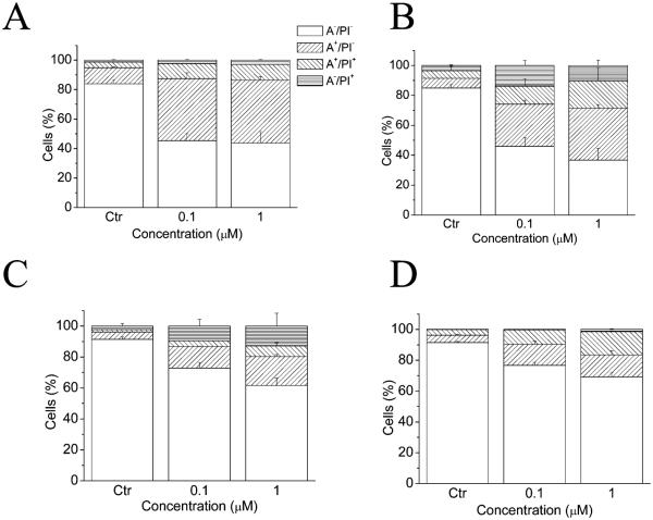
Figure 5.
Flow cytometric analysis of apoptotic cells after treatment of HeLa (A), Jurkat (B), RS4;11 (C), and K562 (D) cells with 5f at the indicated concentrations after incubation for 24 h. The cells were harvested and labeled with annexin-V-FITC and PI and analyzed by flow cytometry. Data are represented as the mean ± SEM of three independent experiments.

