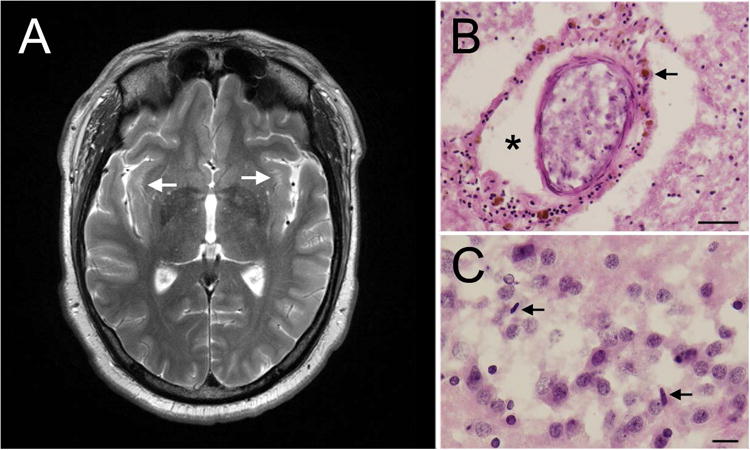
Figure 1.

Imaging and pathology. A, Bilateral white matter hyperintensities in the insula. B, Intraparenchymal blood vessel with perivascular clearing (asterisk) and hemosiderin accumulates (arrowhead) (hematoxylin-eosin; scale bar= 100 μm). C, Scattered rod-shaped microglia seen in the dentate gyrus. Arrowheads point to microglial nuclei (hematoxylin-eosin; scale bar = 25 μm).
