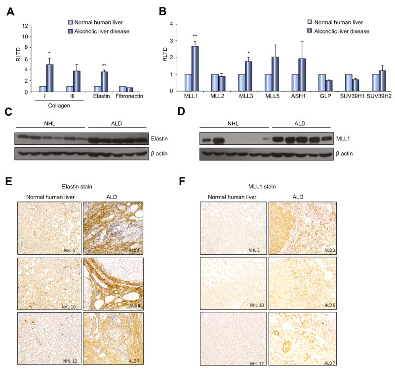
Figure 3. ALD is associated with an increase in MLL1 and elastin expression.
(A) mRNA levels of extracellular components collagen I, III, elastin and fibronectin were quantified by qPCR in six separate preparations of normal human liver or alcoholic liver disease explant. (B) Histone lysine methylatransferases were quantified by qPCR as in (A). Error bars in relevant panels represent mean values ± standard error of the mean (SEM). *p<0.05; **p<0.005. (C-D) Thirty μg whole cell protein from six normal human liver (NHL) or five alcoholic liver disease explanted tissue samples were immunoblotted for MLL1, elastin and β-actin. (E) Representative sections showing MLL1 and elastin immunochistochemical staining in either normal human liver or ALD explanted liver.

