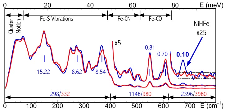
Figure 3.
NRVS-derived PVDOS for DvMF NiR-H (blue) and NiR-D (red) (Ogata, Kramer et al., 2015 ▸). The middle blue labels indicate the signal level (cts/s) for different vibrational features for NiR-H. The bottom labels show the total time (s) measured for each energy regions for NiR-H (blue) and NiR-D (red).

