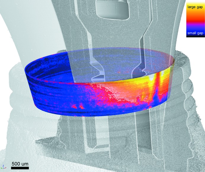
Figure 2.
Exemplary three-dimensional visualization of an IAC microgap map (coloured) superimposed on the CT scan of the dental implant, pointing out the orientation of the latter. Here the implant is a NobelActive (Nobel Biocare, Switzerland) under 333 N cyclic load. The IAC map is calculated according to Zabler et al. (2012 ▸); the look-up table is comparable with Fig. 6 ▸.

