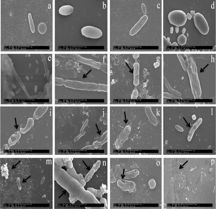
Figure 3.
Representative SEM images of ENPs and metal salts effect on AS microbial cells in relation to control after 60 days exposure. (a–e) Intact microbial cells in control; (f, h) metal salts distorts and shrinks microbial cell; (g) perforations of cells by metals salts; (i, j) cell wall perforation by ENPs; (k) selective adsorption of ENPs to cells; (l) selective cell degenerated by ENPs ions or ROS; (m) selective adsorption to cell and aggregation of ENPs; (n) sheathed and unsheathed cell damage by ENPs; (o) cell wall perforations by ENPs; (p) ENP dissolves cell wall and sheath.

