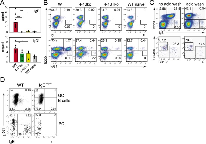
Fig 1. IL-4/IL-13 secreted by CD4+ T cells is necessary for the IgE and IgG1 response.
Serum (A) and mesenteric LN (B and C) of WT, IL-4/IL-13 double deficient mice (4-13ko), or T cell-specific conditional IL-4/IL-13-deficient mice (4-13Tko) were collected from naïve mice or on day 12 after Nb infection. (A) Serum ELISA for total IgE and IgG1. Bars show the mean of individual mice (dots). (B) Cells were washed with acidic buffer to remove cytophilic IgE and permeabilized before staining. Dot plots are gated on live cells as indicated in S8 Fig and show the frequency of B cells (B220+) or B220− cells expressing either IgE or IgG1. (C) Surface staining of B220 and IgE before (left plots) or after (right plots) acid wash in dot plots gated as indicated in S8 Fig. The upper dot plots demonstrate the reduction of cytophilic IgE after wash with acidic buffer on B220+ but not on B220– cells. The lower plots are gated on B220–IgEhi cells and show basophils (CD138–CD49b+) and plasma cells (CD138+CD49b−). (D) Intracellular staining for IgG1 and IgE after acid wash in GC B cells (B220+CD38−GL-7+) and plasma cells (PC, B220loCD138+) from LN on day 10 after secondary Nb infection of BALB/c (WT) and IgE-deficient (IgE−/−) mice gated as indicated in S9 Fig. *p < 0.05, ***p < 0.001 by Student’s t test. Data are from at least four mice and at least two independent experiments.

